In our everyday lives, we all process and recognize hundreds of different objects (colors, shapes, animals, faces…), and although we may not think about it, developing this ability is actually an intricate learning process.
When a young child sees a dog for the first time, they don’t know it’s a dog until someone tells them. They also have no idea that they will eventually encounter many other dog breeds that look completely different from this particular one, but that are still undeniably dogs (and definitely not cats or zebras!). Developing and adjusting such a recognition ability into an accurate and reliable sense of what is and isn’t a dog requires them to go through a series of necessary trials and errors.
To draw a parallel with the example above, although we may initially believe that all dogs look the same, as we witness more breeds of dogs, we gradually alter the mental picture we have of dogs to encompass their diversity. The more dogs we see, the more accurate and generalizable our ability to recognize them becomes. Eventually we learn to differentiate between the common features that dogs share (e.g. dogs have four legs), from those specific to their sub-groups (e.g. Dalmatians are white with black spots).
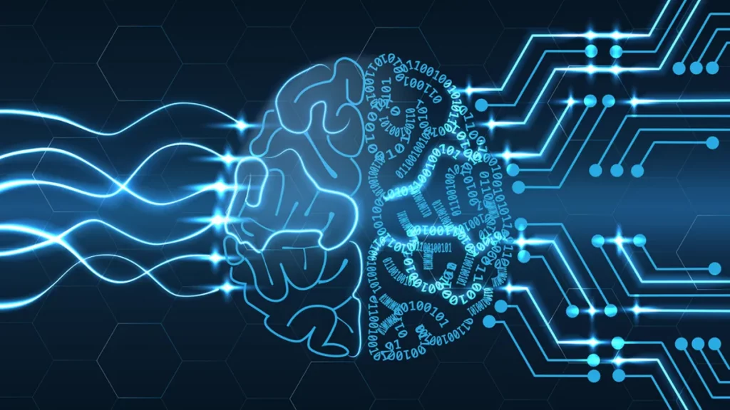
Much like people, machines can be taught object recognition by mimicking the learning process of the human brain. This process, called Deep Learning, is an application of artificial intelligence that, although designed to learn through a specific set of data at first, can continue to learn on its own and improve from experience, without being explicitly programmed to do so.
Deep learning becomes more accurate with time. This is because each analysis provides feedback on the accuracy of its performance, allowing it to learn from its mistakes. While the initial algorithm does not change, the code’s internal weights and biases are adjusted with each trial and error, improving its odds of answering correctly the next time. With experience, the algorithm develops the ability– like humans – to recognize patterns of similarity between objects and group them into categories (like dogs) and sub-categories (like Dalmations or Dachshunds).
Because of its ability to automatically categorize and classify, deep learning holds great potential in both research and clinical fields. For example, deep learning can be a powerful tool in improving diagnostic accuracy, as it has the ability to discern subtle abnormalities in medical results such as radiological images.
In magnetic resonance imaging (MRI) – a widely used non-invasive imaging technique – deep learning can be helpful in differentiating scans of patients and healthy individuals by comparing their anatomical and/or functional features. This is possible when one or more MRI-detected features of the brain correlate strongly with a specific disease or condition. Even if present across an array of diseases, a feature can still be useful if its characteristics (e.g., volume, shape, density…) differ significantly between diseases. If so, the feature can be accepted as a biomarker; a naturally occurring characteristic that can be used to identify a particular pathological and/or physiological condition.
An example of an MRI-detected feature that is an established biomarker of brain lesions is white matter hyperintensity (WMH). While it is widely accepted that WMH is a frequent finding in the aging population, its association with age-related dementia is being increasingly recognized.
More studies are now seeking to assess whether there are reliable differences in the characteristics of WMH in the brain (i.e., volume, shape, spatial distribution) in different types of neurodegenerative diseases such as Alzheimer’s and Parkinson’s disease. If so, this would imply that deep learning could be used to reliably diagnose different types of dementias by looking for specific characteristics of WMH. This becomes especially useful when considering how difficult it can be for doctors to distinguish between neurodegenerative diseases that have very similar symptoms, as is the case for many of the dementias, especially in the early development stages of the disease. Characterizing how WMH features vary across the age-related dementias can help us design a deep learning algorithm that may contribute to an earlier and more accurate diagnosis of these neurodegenerative diseases.
Although deep learning remains a novel and developing field, this example underlines how deep learning can bring important contributions to both research and clinical practice. In the same way that doctors and radiologists learn to recognize, categorize, and diagnose with years of experience, deep learning has the potential to do the same thing, only much much faster.

Ikrame is a Master's student in Biomedical Engineering at the Université de Montréal. Her current work centres around investigating the relationship between MRI- detected cerebrovascular pathology, cerebrovascular architecture, and age-related dementias. Ikrame's passion for scientific research extends beyond bench work, as scientific communication has been integral to her research involvements. Making science accessible to the general audience is crucial, and she believes that knowledge translation is key to achieving this goal and hopes to contribute to this effort.