[Cet article est en anglais] Last summer, QBIN launched the first trial of a new program designed to engage cégep students in bio-imaging research and help graduate students and postdocs gain valuable mentorship experience. Three PhD students from the Integrated Program in Neuroscience at McGill University were paired with two cégep students from Dawson College to complete a 10-week research internship. The pilot was a great success and we plan to expand the program to include more mentors and interns from across the province in summers to come! Read on to learn more about the program from the perspective of our first three QBIN mentors, Aurelie Bussy, Stephanie Tullo, and Isabelle Arseneau-Bruneau!
Each PhD student was in charge of proposing a project and mentoring their two cégep students to complete the project from June to August 2022 and were compensated for their time. For the cégep students, their internships culminated in a poster presentation explaining their projects to their peers, much like a miniature research conference.
The pilot was a great success and we plan to expand the program to include more mentors and interns from across the province in summers to come! Read on to learn more about the program from the perspective of our first three QBIN mentors, Aurelie Bussy, Stephanie Tullo, and Isabelle Arseneau-Bruneau.
Stephanie Tullo, PhD candidate working at the Computational Brain Anatomy laboratory at the Douglas Mental Health University Institute under the supervision of Prof. Mallar Chakravarty
It was a pleasure to participate in this pilot mentorship program brought about by QBIN and Dawson. This opportunity allowed me to gain mentorship experience, specifically in terms of developing a suitable research project, devising a plan and creating a feasible timeline, communicating complex concepts into simple, easy to digest material, and hosting weekly meetings to assess their progress. Given the students’ education level and their limited experience in research, an important skill that I developed as part of this program was that by tailoring the material to their current knowledge, I gained a broader basic understanding of the techniques and methods that I use on a daily basis. Another great moment of this program was watching the students learn challenging concepts and techniques and now being able to explain the work to their peers and seeing the pride they had when talking about the work they completed over the summer. I would most definitely recommend this program to other graduate students, and I would participate again next summer.
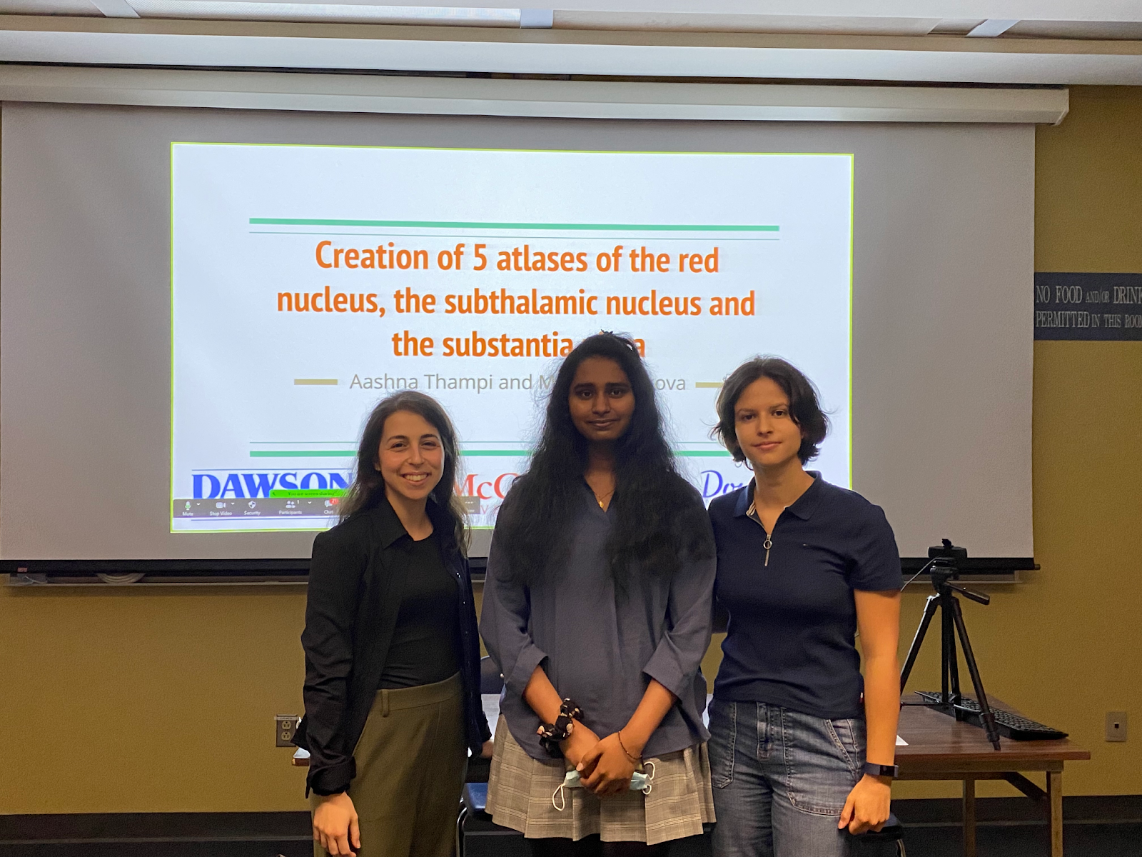
About the project
As part of this program, the students manually delineated (segmented) three key structures in Parkinson’s disease (the substantia nigra, the subthalamic nucleus, and the red nucleus) on 5 high-resolution brain images using various MRI contrast images (T1-weighted, T2-weighted, and T1-weighted/T2-weighted ratio images) to better visualize these small structures. The ultimate goal is for these new atlases to be implemented as part of the automatic MAGeT-Brain segmentation pipeline. With this work, the students gained experience in the neuroanatomy of the brain and in learning about MRI. By the end of the program, the students were able to identify various subcortical structures of the brain (especially the basal ganglia system) and be familiar with various types of MRI images and contrasts, understanding how to interpret the signal from each type of image, in terms of the biology of the brain and which method is most appropriate for different types of research questions.
Aurelie Bussy, PhD candidate working at the Computational Brain Anatomy laboratory at the Douglas Mental Health University Institute under the supervision of Prof. Mallar Chakravarty
I really enjoyed being part of this amazing project! I believe it has definitely given a lot of insights to the students about what is the world of research and how a research lab works. Through this summer, I really liked sharing my knowledge with them and gaining mentorship experience. It helped me to be better organized and find simple ways to explain complex things. I would definitely recommend this mentorship program for both cégep and PhD students since it really provides great outcomes for all!
About the project
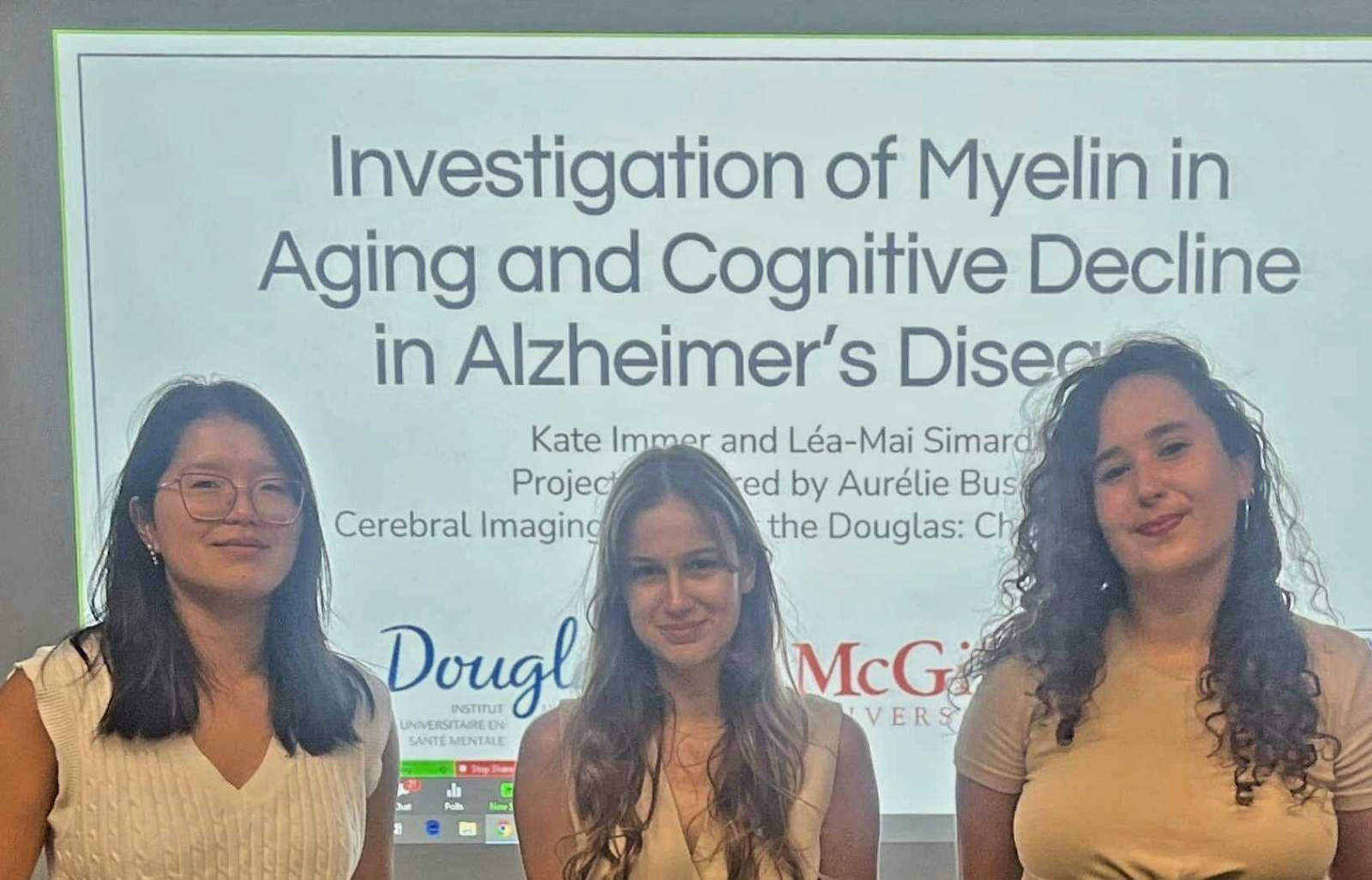
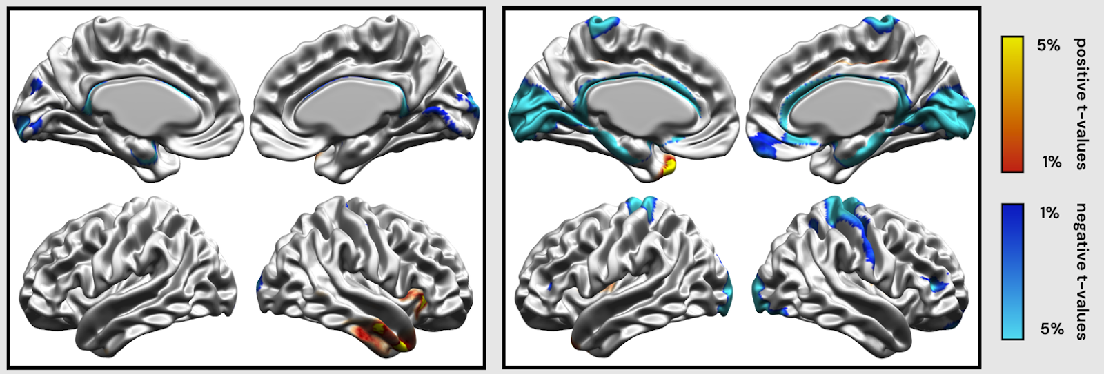
The project that I proposed was to investigate the myelin in aging and cognitive decline in Alzheimer’s disease progression. Indeed, as the life expectancy of the Canadian population increases, one consequence is the increased prevalence of neurodegenerative disorders such as Alzheimer’s disease. To study human brains, magnetic resonance imaging (MRI) is a powerful imaging technique with which we can measure potential biomarkers such as myelination. Myelin is an important component of the brain, and previous studies have highlighted myelin impairment in healthy aging, and further myelin reduction in Alzheimer’s disease. Therefore, in this project, we sought to use an MRI biomarker of myelin to characterize individuals across the disease spectrum: from healthy aging individuals, to individuals with mild cognitive impairment or AD dementia.
The students performed the preprocessing of MRI brain scans of 600 individuals at various stages of cognitive decline. They learned brain neuroanatomy, how to rate the quality of brain scans by looking for potential image artifacts and the basic physics behind the MRI technology. They ran various processing pipelines in order to preprocess the images, create T1w/T2w ratio and extract cortical vertex-wise information. To perform the image processing and the statistical analyses, the students learned coding skills such as bash and R. The students also investigated how the myelin was evolving with age and cognition by doing statistical analyses.
Isabelle Arseneau-Bruneau, PhD candidate working at the Zatorre Laboratory (Auditory Cognitive Neuroscience) at the Montreal Neurological Institute under the supervision of Prof. Robert Zatorre

I very much enjoyed and recommend the QBIN Mentorship Program! I hope that it was as useful for the cégep students that it was for me as a mentor. I believe that it contributed to the development of solid fundamental research skills, gave the students a taste of laboratory environments, and generated an opportunity for continuous learning from college through university. When I saw one of the students during their poster presentation at the beginning of October, I was glad to learn that some of the knowledge acquired this summer was useful in their coursework this fall. On my side, I gained experience teaching experimental procedures, explaining complex content in simpler ways, establishing expectations and frameworks for our team that were useful to give direction to the various aspects of the project. As our research faced considerable challenges (notably technical), I definitely developed some project management skills to optimize our time at the lab. Further, research can involve many necessary but tedious tasks, so it is much more motivating and efficient to face those as a team! Overall, I salute this initiative from the QBIN. One of the students even continued to work for our lab as a research assistant this year, in parallel to his first year at Concordia University.
About the project
Our original project was to compare the frequency following response (FFR) of highly trained pianists and non-musicians while they play on a digital organ and while they hear the same sounds passively, with different placements of the electroencephalography (EEG) electrodes on the scalp (2×2 Design). The FFR is a neural response to auditory stimuli (speech or music) generated by multiple structures of the brain. It provides insights into how accurately the brain encodes key features of sounds, as it reproduces them in the oscillatory activity captured with EEG. Different experimental parameters, such as the electrode montage, affect FFR results as they capture different anatomical contributors to the signal (Coffey et al., 2019). The students had to explore the literature and collect a small sample of EEG data to find which electrode placement could be more optimal for an FFR signal susceptible to involve more cortical (rather than only subcortical) contributions. Within the timeframe of the internship they completed the review of literature and pilot tests.
The students also completed a large variety of tasks associated with auditory cognitive neuroscience research. They first acquired knowledge about the frequency following response and learned to identify the anatomical structures of the auditory pathway. From their review of literature on electrode placements, they produced a table that summarized the findings. This exercise developed their synthesizing skills, to present content in concise but precise manners, which was useful when the students later contributed to research documentation (protocols, run-sheet, etc.) Regarding practical skills, the students completed a Matlab course and then created auditory stimuli with this programming language. They performed audiometry screening, learned about the functioning of EEG, positioned electrodes on the head of participants, took impedance measurements, collected pilot data for the organ active vs passive conditions (EEG) and for behavioral auditory tasks. Last but not least, the students were a huge help to organize the research equipment and label various parts of the experimental set-up, an exercise that was quite helpful when trouble-shooting!

Thank you and congratulations to all of our first mentors and interns!
If you would like to learn more about the program or help us expand it to your city in the future, please get in touch at qbincomms@gmail.com